Josep Brugada (Barcelona, Spain), Carlo Pappone (Milan, Italy) and others report in Circulation: Arrhythmia and Electrophysiology that ablation of abnormal epicardial substrate-identified after flecainide testing-in patients with Brugada syndrome can eliminate the phenotype expression of the syndrome. Although they state that further study is warranted, Brugada et al claim that their results suggest “major physiopathological and clinical implications” for the management of Brugada syndrome.
The authors note that a recent study has suggested that “the electrophysiological substrate for the Brugada syndrome is delayed depolarisation exclusively over the anterior aspect of the right ventricular outflow tract epicardium and that catheter ablation of this abnormal area may prevent ventricular tachycardia/fibrillation” in patients with the syndrome. However, they add that “it is unknown” if there is a relationship between the presence, location, and extent of the epicardial substrate abnormalities and typical Brugada syndrome ECG pattern. “In addition, many other issues such as methodology for substrate identification and elimination and testing of acute and mid-term results remain to be clarified,” Brugada (Arrhythmia Section, Cardiology Department, Thorax Institute, Hospital Clinic and IDIBAPS, Barcelona, Spain) et al write.
Therefore, in the present study, the authors sought to “systemically report” the methodology, results, and complications of epicardial radiofrequency ablation in consecutive patients with Brugada syndrome, stating: “Particular attention was focused on endocardial/epicardial mapping and flecainide testing to characterise and establish the most appropriate target sites for successful radiofrequency ablation and elimination of both ECG Brugada syndrome pattern and inducibility of ventricular tachycardia/fibrillation.”
In the study, 14 consecutive patients with documented spontaneous and/or flecainide-induced type I Brugada syndrome ECG pattern and symptoms of ventricular arrhythmias, high vulnerability for ventricular arrhythmia induction, and who had an implantable cardioverter defibrillator (ICD) underwent high-density detailed endocardial and epicardial electroanatomical mapping and ablation with a 3D mapping system (Carto 3, Biosense Webster) before and after flecainide infusion. Brugada et al comment: “Once epicardial low voltage areas were identified and quantified after flecadinide, all abnormal electrograms inside these areas were tagged in order to be targeted for radiofrequency delivery.” They note that radiofrequency was delivered using an externally irrigated 3.5mm tip mapping/ablation catheter (Thermocool, Navistar, Biosense Webster).
All patients had evidence of abnormal epicardial electronanatomic voltage maps-characterised by low voltage (<1.5mV) areas and low frequency, slow late potentials, variable for extension and distribution before and after flecainide. Brugada et al report that, overall, both the median baseline low voltage area and the median abnormal epicardium electrogram area significantly increased after flecainide infusion and comment: “Interestingly, there was a relationship between ECG pattern changes and low voltage area. The wider the low voltage area (<1.5mV), the higher the ST segment elevation and covered type appearance.”
They state that after a median radiofrequency application duration of 23.8 minutes, local abnormal ventricular electrograms “completely disappeared” in low voltage areas and were replaced by residual dense scar areas (<0.5mV) of 25.9cm. The authors add: “After radiofrequency ablation, all patients became non-inducible during programmed electrical stimulation using up to three extrastimuli. ECG did not show any change suggesting Brugada ECG pattern after flecainide.” Furthermore, Brugada et al comment, epicardial electroanatomic voltage remap confirmed “complete elimination of abnormal electrogram in low-voltage areas” in all patients. According to the authors, this substrate elimination was associated with the absence of Brugada syndrome ECG pattern. They state that at the five-month follow-up point: “ECG remained normal despite flecainide testing and ICD did not show arrhythmia events.”
Concluding, Brugada et al write that their study provides “new insights” into the mechanism, prevention, and treatment of Brugada syndrome. They add: “Although larger studies with longer follow-up are required, our results have major physiopathological and clinical implications as they provide new important information leading to a potential new definitive elimination of the phenotypic manifestations of Brugada syndrome.”
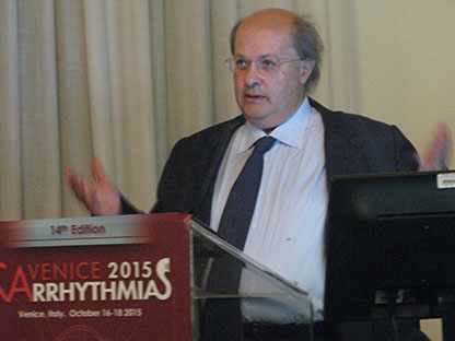
However, after study author Carlo Pappone (Policlinico San Donato, University of Milan, Department of Arrhythmology, Electrophysiology & Cardiac Pacing, Milan, Italy) presented the results at Venice Arrhythmias (16–18 October, Venice, Italy), a delegate questioned the findings of the study-he said: “I think what you have discovered is not necessarily the Brugada syndrome. If you take anyone with abnormal ECG, even if mildly abnormal, and you give them drugs such as flecainide, the ECG will become much more dramatically abnormal. The more abnormal it is, the more abnormal it becomes with these drugs. So I think what you have discovered that many people have patchy layered scar on the epicardium and I would bet that if we give these drugs in other settings, you will see similar types of local ECG changes and dramatic ECG changes.”
Pappone told Cardiac Rhythm News that he and Josep Brugada “strongly disagreed” with the delegate’s interpretation of their work. He says: “There are many publications in the literature coming from different groups around the world showing that the use of flecainide-or ajmaline-is highly sensitive and specific for the identification of patients with Brugada syndrome. In some of these patients, the presence of a pathological mutation in the sodium channel is used as a gold standard test for the diagnosis. Studies have shown that in patients with the Brugada mutation, changes in ECG are systematic after drug administration but no significant changes are produced with the drug in patients without the mutation-except for a slight PR and QRS prolongation, as would be expected with a class I antiarrhythmic drug. Furthermore, administration of flecainide or ajmaline in patients with isolated right bundle branch block or with clear structural heart disease-such as arrhythmogenic right ventricular cardiomyopathy-in the vast majority of cases does not produce ST segment elevation, but prolongation of the activation time in the right ventricle or in some extreme cases, prolongation of the duration of the epsilon wave. Also, an ajmaline test has been used for decades in the diagnosis of conduction disturbances and changes similar to Brugada pattern have not been documented in this setting. Therefore, the literature and our own experience indicates that the administration of flecainide or ajmaline in patients without a Brugada syndrome background does not produce changes similar to the ones induced in Brugada syndrome patients and strongly supports the idea that the changes observed after drug administration in the local and surface ECG are very specific for patients with Brugada syndrome.”


