Syed Rafay A Sabzwari and Dhanunjaya Lakkireddy (Kansas City, USA) review the importance of obstructive sleep apnoea (OSA) in electrophysiology and cardiology at large, as a secondary impact to an underserved area of healthcare, which also provides practical implications for the cardiology practice in general.
Obstructive sleep apnoea is a common sleep related breathing disorder with an estimated prevalence of 4% in men and 2% in women.1 Due to partial or complete collapse of the upper airway during sleep, it is characterised by repetitive episodes of reduced respiratory flow leading to intermittent hypercapnia, hypoxemia and arousals to re-establish airway patency. Seemingly a primary respiratory and airway disorder, the complex pathophysiologic mechanisms involved have a greater negative systemic impact than what is generally perceived. Its effect on the heart is a significant part of this systemic milieu. In our own experience less than 20% of patients are probed about symptoms of OSA in cardiology clinics. This shows that not only does this relationship remain undiagnosed and untreated in 80% of apnoea sufferers but whenever it is diagnosed it is underdiagnosed and undertreated.
More than 90% of cardiovascular conditions and comorbidities are affected by OSA. These include drug-resistant hypertension (83%), congestive heart failure (76%), diabetes mellitus type II (72%), stroke (63%) and coronary artery disease (57%).2 Even more, it affects cardiac arrhythmias (58%)2 including but not limited to atrial fibrillation, sinus node dysfunction, ventricular ectopy, ventricular tachycardia and sudden cardiac death. Intermittent hypoxia and hypercapnia stimulates chemoreceptors, baroreceptors, pulmonary afferents, coupled with reduced venous return to the heart causes increased sympathetic activity directly affecting heart rate and blood pressure.3 The respiratory acidosis and autonomic nervous system fluctuations together result in triggered automaticity and re-entrant mechanisms. In addition, the amplified negative intrathoracic pressures lead to increased juxtacardiac and transmural pressures on the thin-walled atria playing an important role in atrial arrhythmogenesis.

Risk of atrial fibrillation is four times higher in patients with OSA independent of obesity, age, hypertension and heart failure or other confounding variables. Conversely, 49% of patients (3.5 million US population) of atrial fibrillation patients have OSA.2 This has been supported by multiple studies. In one study with 400 patients with moderate to severe OSA, 24-hour holter monitoring showed atrial fibrillation in 3% of patients, which is three times higher the general prevalence.4 Preceding hypopneas before paroxysms of atrial fibrillation shown in a case cross design supports an immediate causal temporal relationship.5 Even more significant is the increasing data implicating OSA as a risk factor for recurrence of atrial fibrillation after ablation or cardioversion. In a prospective study with 720 patients by Neilan et al, the presence of OSA (hazard ratio 2.79, CI 1.97 to 3.94, p<0.0001) and untreated OSA (hazard ratio 1.61, CI 1.35 to 1.92, p<0.001) were highly associated with atrial fibrillation recurrence.6 The same study showed reduction in blood pressure, atrial size and ventricular mass in patients treated for OSA. Similarly, a meta-analysis showed that in patients undergoing pulmonary vein isolation or medical management for atrial fibrillation, there was a 42% relative risk reduction in atrial fibrillation recurrence with the use of continuous positive airway pressure.7 This means that atrial fibrillation treatment outcomes can continue to be in jeopardy if OSA is not adequately addressed and should not just be taken as a causative factor for atrial fibrillation but a continued insult in the natural progression of atrial fibrillation.
Furthermore, tachycardia-bradycardia phenomenon often seen in atrial fibrillation patients may also be seen with OSA. Enhanced parasympathetic tone from vagotonic activation of carotid body has been anecdotally thought to cause sinus and atrioventricular blocks; however, multicentre epidemiologic data8 do not reflect this association any more than that observed in patients without OSA. Nonetheless, ventricular tachyarrhythmias and QT prolongation in OSA could be a plausible additional predisposing factor.9 An observational study with 228 patients showed a higher prevalence of non-sustained ventricular tachycardia (5.3 vs. 1.2%) and complex ventricular ectopy (25 vs. 14.5%) including bigeminy and trigeminy.10 Moreover, sudden cardiac death caused by fatal ventricular arrhythmia, although it has limited data, has also been associated with OSA. In a large, observational study of over 10,000 patients of whom 80% had OSA, multiple OSA severity indicators were associated with increased risk of incident sudden cardiac death.11
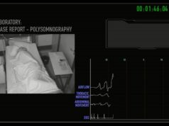
An attended overnight polysomnography recording has been the standard diagnostic test for OSA. However, polysomnography has been unpopular and perceived as a cumbersome process when persuading patients to undergo testing, letalone the costly labour, equipment requirements, long drawn out process with delayed results and the need for designated centres with already long waiting times. In the effort to overcome this diagnostic hurdle, multiple ambulatory sleep study devices have been experimentally developed and their accuracies have been compared to polysomnography. Amongst them, devices using peripheral arterial tonometry (PAT) such as the WatchPAT device (Itamar Medical) has undergone scrutiny by various studies and has therefore gained popularity.
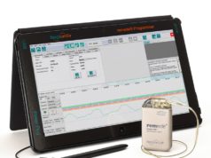
WatchPAT is a unique, ambulatory sleep study device worn on the wrist that uses peripheral arterial tonometry together with pulse oximetry and actigraphy to measure respiratory disturbances. Changes in sympathetic tone of the vasculature corresponding to episodes of apnoea and hypopnea are collected by the device and correlated with pulse oximetry readings formulating results using a predeveloped automated algorithm thereby offering a simpler, reproducible, accurate, much more affordable alternative to polysomnography by an in-house cardiology team offering greater patient flow and, that too, in the setting of patient’s own house. Other advantageous features include its ability to measure body position for assessing position related OSA, measuring total sleep time instead of total sleep duration, providing rapid eye movement (REM) related OSA diagnosis and a detailed sleep architecture for more accurate diagnosis of the severity of OSA (mild, moderate, severe). Multiple studies with small sample sizes have identified high correlation of sleep indexes such as respiratory disturbance index (RDI), apnoea-hypopnea index (AHI) and oxygen desaturation index (ODI) to those measured by formal polysomnography.7 A meta-analysis reviewed 14 studies encompassing 909 participants and correlated these indexes between peripheral arterial tonometry and polysomnography; RDI correlation 0.879 (CI 0.849-0.904, P<0.001); AHI correlation 0.893 (0.857-0.920, p<0.001); ODI correlation 0.942 (0.894-0.969, p<0.001).7
Nevertheless, peripheral arterial tonometry devices are less sensitive than polysomnography, therefore patients with a high clinical suspicion and negative results should be evaluated through formal polysomnography before a diagnosis of OSA is rejected. Also its use is contraindicated in patients with moderate to severe pulmonary disease, neuromuscular disease, congestive heart failure, central sleep apnoea and periodic limb movement disorder.7 Certain patient groups with vascular disease which might impair the arterial tone like diabetes mellitus, vasculitis will also not give accurate results, hence were excluded from the studies.7 Despite the fact that advancing age causes reduced compliance, the inclusion of elderly patients in studies has shown no difference in peripheral arterial tonometry compared to polysomnography data in the geriatric population.7 The ability of the peripheral arterial tonometry devices in accurately recording the sleep apnoea monitors may be limited in patients who are in persistent atrial fibrillation as a significant amount of the data generated by these devices is dependent on the arterial tone which may be affected by atrial fibrillation.
One cannot claim mission accomplished by merely establishing the diagnosis and starting continuous positive airway pressure therapy. One has to remember that OSA is a dynamic disease that continues to evolve with patient’s age, biophysical profile, comorbidities, medications, stress levels etc. It brings the need for periodic monitoring and adjustment of continuous positive airway pressure settings with changes in patient’s conditions. With more than 50% patients with OSA being unable to use their continuous positive airway pressure machine properly, the issue of compliance makes itself very apparent. This challenge is also evident in the fact that more than 90% of patients did not have a follow-up visit with their sleep specialist after the initial visit. More than 50% have been using the same continuous positive airway pressure settings since their sleep study and more than 50% patients had a change in their BMI or new diagnosis of atrial fibrillation, ventricular tachycardia or heart failure since their OSA diagnosis. Due to the ease and cost-effectiveness of portable home monitoring, it offers a reasonable solution in this area. Studies have shown that outpatient’s in-home auto-titration of positive airway pressure saw continuous positive airway pressure adherence and clinical outcomes similar to polysomnography.12 In another study, in-home peripheral arterial tonometry devices accurately identified participants with residual moderate to severe sleep disordered breathing while using continuous positive airway pressure.13
Management of OSA requires a multidisciplinary approach and the burgeoning burden of OSA on the heart requires a proactive forefront role to be played by cardiologists especially in the social context where patients are more likely to abide by the instructions and recommendations of a cardiologist than another physician. Using the peripheral arterial tonometry device, cardiologists can be at the centre of the communication circle between the primary care, sleep specialists and pulmonologists. A good working model could have one or two sleep specialists on speed dial for referrals. Each cardiology satellite clinic could have two to three peripheral arterial tonometry devices that are given out to patients, mailed back to the clinic after use, cleaned and restocked for reuse. The downloaded data from the device can be electronically transmitted to the sleep specialist for interpretation eventually having results in the EMR system with an action plan. This strategy offers success for both physicians and patients and reflects great partnership in disease control.l
To summarise, OSA is a debilitating, chronic condition with a great impact on cardiovascular conditions and if left untreated can compromise
treatment of common cardiovascular comorbidities including arrhythmias as well as overall patient care and cost. Its prevalence is expected to continue escalating given the obesity epidemic.7 Therefore, cardiologists need to become more aware and proactive in coordinating early diagnosis, treatment and continued treatment titration of OSA with the utilisation of easy to implement and practically integrate ambulatory sleep study devices and create an exemplary partnership in disease prevention and propagation. A multi-specialty approach to a disease process that has such profound impact on cardiovascular health needs a full engagement of the treating cardiologist along with the sleep specialist.
References:
- Young T et al, N Engl J Med 1993;328(17):1230–1235
- Seet E et al, Anesthesiol Clin 2010 Jun;28(2):199–215. doi: 10.1016/j.anclin.2010.02.002
- Friedman O, Logan AG, Am J Hypertens 2009;22:474
- Guilleminault C et al, Am J Cardiol 1983;52:490
- Monahan K et al, J Am Coll Cardiol 2009;54:1797
- Neilan et al, J Am Heart Assoc 2013;2:e000421
- Yalamanchali et al, JAMA Otolaryngol Head Neck Surg 2013;5338
- Mehra R et al, Am J Respir Crit Care Med 2010;182:826
- Shamsuzzaman AS et al, Sleep 2015;38:1113
- Mehra R et al, Am J Respir Crit Care Med 2006;173:910
- Gami AS, et al, J Am Coll Cardiol 2013;62:610
- Berry et al, Sleep 2008;31(10):1423–31
- Pittman et al, Sleep Breath 2006 10:123–131
Syed Rafay A Sabzwari and Dhanunjaya Lakkireddy are with the Section of Electrophysiology, University of Kansas Hospital, Kansas City, USA. Lakkireddy receives a speaker’s honorarium from Itamar Medical


