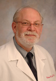
Roberto M Lang, professor of Medicine, University of Chicago Medical Center, Chicago, USA, is a pioneer in the development of 3D echocardiography. Lang, who is past president of the American Society of Echocardiography, spoke to Cardiovascular News about the use of 3D echocardiography and the future of the technique.
You were a pioneer in the development of 3D echocardiography, which has now been around for several years. How has it changed clinical practice in the cardiovascular field?
Three-dimensional echocardiography has been around for 30 years, but only in the last eight to 10 years that it has become a real-time technique. You place the transducer on the patient’s chest and see a 3D picture on the screen without the need for any reconstruction.
Three-dimensional echo has two ways to obtain the information. One is from the thorax, called transthoracic. The second one is when the patient ‘swallows’ a gastroscope so we can obtain a transoesophageal image. Transoesophageal 3D echo has two major roles. It can display the mitral valve in a way that is similar to the way a surgeon sees it when he opens the patient’s chest in the operating room. It has played a major role in preparing patients for mitral valve surgery and, in particular, for mitral valve repair. It allows the surgeon to know what he is going to find, and therefore what he will need to do during the surgery. The second aspect is that there are a lot of interventions that are now being done in the cath lab for structural heart disease such as atrial and ventricular septal defects, clip on the mitral valve, implantation of occluder devices to treat perivalvular leaks, etc. All these interventions are being done with the guidance of three-dimensional echo. In the past, without 3D echo, they were not possible.
In transthoracic echo, the major characteristic is that 3D echo allows the physician to quantify the pumping ability of the heart with much more accuracy. Transthoracic 3D echo is useful for the quantification of left ventricular volumes and function.
In transcatheter aortic valve implantation, how can 3D echocardiography facilitate diagnosis of aortic stenosis and help in planning the procedure?
There are several studies, particularly from Europe, that have incorporated 3D echo into the quantification of the aortic valve area. That is quite new. The most important role that 3D echo has played is in mitral valve intervention, and now also for TAVI, in the guidance of these types of procedures. With the advent of 3D colour Doppler, it is also possible to use 3D echo to quantify the stability of the valvular leaks.
What is the role of real-time 3D transoesophageal echocardiography for screening and patient selection for left atrial appendage closure?
For the left atrial appendage closure, 3D echo can be very good to assess the size of the left atrial appendage mouth, so you know what size of device you need to use.
How do you see the use of echocardiography in the future?
Many times, in order to acquire the 3D echo image, you need to do it in four consecutive bits. For example, we cannot obtain pristine images in people who have arrhythmias. As computers improve, we will be able do all these acquisitions in one bit. The other thing that is going to happen is the miniaturisation of the probe that we put down the patient’s throat. It is going to become smaller, and we will maybe be able to place it through the patient’s nose, without the need for anaesthesia.
We will also be able to have a 3D colour feature to look at the leaks. However, there is still a lot of improvement to be done in this area. Ideally, companies will be able to devise systems which will acquire images easily and more objectively.
How do you see echocardiography versus cardiac MRI? Are they complementary tests?
They are totally complementary techniques. For a while, they were considered competitor techniques. A busy hospital can probably do 60–70 echocardiograms a day, but only five or six MRIs. So it is not really a competition. MRI provides information that echo cannot provide; for example, whether a particular portion of the muscle is scarred or not. Also, perfusion by an open vessel is something that MRI can provide with good information.
Developments in heart modelling have also been improving cardiac care and helping in the training of cardiovascular interventionists. How do you integrate ultrasound in cardiac modelling?
With cardiac CT, physicians can press a button and it will totally and automatically isolate and separate the four different chambers of the heart. It will calculate volumes and how much these chambers pump. All four chambers of the heart can be done automatically, and there is no variability. The same idea is being developed with 3D echo. The objective is to be able to quantify each of these portions of the heart chambers automatically and take away any variability.
In your opinion, what are the most exciting imaging technologies available on the market at the moment?
We do a lot of 3D echo imaging. We work closely with Philips because they have been a pioneer in 3D echo. Also, because Philips was the only company that had a transoesophageal probe. Now GE also has it. In terms of 3D Philips is very advanced. We use a 3D echo machine using a matrix array probe.
What advice would you give to young cardiologists who wish to work with 3D cardiac ultrasound?
Echocardiography is a technique that was initially developed in Sweden about 60 years ago. It is a very mature technique, which is used all around the world. Initially, images were acquired in a single dimension, then after that images were acquired in two dimensions. Recently the technology evolved to three dimensions. If you are a young physician, this is a very good time to hop on the wagon and learn about 3D echocardiography because this technique is here to stay.


