
Details of the first-in-human use of an augmented reality holographic display to aid physician accuracy during cardiac ablation have been published in the Journal of the American College of Cardiology (JACC: Clinical Electrophysiology).
Researchers at Washington University (St Louis, USA) led by Jennifer N Avari Silva, associate professor of pediatrics at the School of Medicine, and Jonathan Silva, associate professor of biomedical engineering in the McKelvey School of Engineering, co-led a team that tested a Microsoft HoloLens headset with custom software during cardiac ablation procedures on patients at St Louis Children’s Hospital (St Louis, USA). Results of the trial were also presented at the International Conference on Human Computer Interaction (July 10 2020), 2020 and published in the IEEE Journal of Translational Engineering in Health and Medicine.
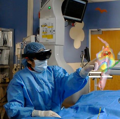
In an ablation procedure using an electroanatomic mapping system (EAMS) a technician controls the catheters while the physician views the images on monitors presented in two different planes. The physician then has to mentally create the image of the heart. Jennifer Silva, also director of pediatric electrophysiology at St. Louis Children’s Hospital, said that standard-of-care technology is antiquated, and the team believed it could do better using an augmented reality system.
Jon Silva and his team of engineers created software for the Microsoft HoloLens headset that converts the data from the catheters fed into the patient’s heart into a geometrical holographic image that hovers over the patient. The headset allows the physician to take control of the procedure by using his or her gaze to guide the controls and to keep hands free and sterile. Their system, Enhanced Electrophysiology Visualization and Interaction System (ĒLVIS), provides a 3D digital image of the patient’s electroanatomic maps that provide a picture of the inside of the heart, which they can measure and manipulate during the procedure.
Jon Silva said that the technology has caught up with the application. The headset allows the user to view their whole environment, including the patient, unlike virtual reality, which take a user completely out of their environment.
“The old headsets were slow to update and made the users sick,” he said. “The development of mobile phones and mobile technology and computer has enabled these kinds of displays and headsets.”
To test the device, two physicians at St Louis Children’s Hospital received a short training session on the device before using it on a total of 16 paediatric patients. During a post-procedure waiting phase, the physicians were given 60 seconds to navigate to each of five target markers within the geometry of the heart, using both the 3D ĒLVIS and the 2D EAMS technology. The physicians were significantly more accurate with the ĒLVIS technology, according to the study team.
“Without the use of the ĒLVIS 3D display, a significant fraction of ablation lesions, 34%, would be made outside of the target area, as opposed to 6% with ĒLVIS 3D display,” Jon Silva said. “We expect that this will improve patient outcomes and potentially reduce the need for repeat procedures.”


