

Benjamin D’Souza, assistant professor of Medicine at the University of Pennsylvania, cardiac electrophysiologist at Penn Presbyterian Medical Center, Philadelphia, USA, discusses the recent developments in robotics in the electrophysiology (EP) lab.
The two main criticisms of the use of robotics in EP I have come across in my discussion with colleagues are longer procedural times, compared with manual use of a catheter, and inferior efficacy. This is largely what I believed coming out of my fellowship at the Hospital of the University of Pennsylvania. During that time, my exposure to the use of robotics was limited, although we performed hundreds of manual cases during those two years.
Due to the lack of exposure and experience in robotics, when I became an attending physician at Penn Presbyterian Medical Center, I did not seriously consider the technology for my practice. Although the system was available for my use, in my first few years at Penn Presbyterian I did no robotic navigation cases and continued to do all cases manually.
However, my opinion on this technology changed. By chance, I observed a robotic ventricular tachycardia (VT) ablation performed by a colleague that I had trained with, and this convinced me to try one of my own. After having a very positive experience I began to incorporate the use of robotics in my VT ablation cases. Over time, with simple workflow changes and further education, my experience with the use of robotics grew, and I was able to accurately and efficiently perform these complex procedures. I currently use the robotic magnetic navigation (RMN, Stereotaxis) system for all of my ventricular ablation cases, and increasingly for other applications I had not previously considered. With RMN I have been able to significantly improve my efficiency, safety, and outcomes.
What is RMN and how does it work?
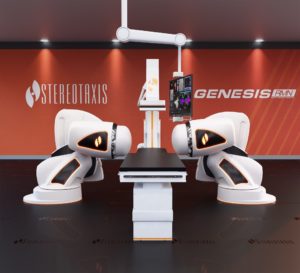
A Robotic Magnetic Navigation system consists of two precise, computer-controlled magnets on robotic arms, a flexible catheter with a small magnet in the tip, and an operating control room. The system creates magnetic fields, which can be finely manipulated to steer catheters directly from their distal tip with an extremely accurate degree of precision and control. Because the catheter is controlled from the distal tip, it is flexible and atraumatic, reducing risk of perforation and improving safety for patients.
Safety first
One of the main reasons I adopted robotic navigation in the EP lab is safety. The clinical benefits of robotic navigation and its use in ablation are well-documented in dozens of peer-reviewed publications, and many other high-volume EP physicians already use this technology. For those who have observed the use of the system and catheter, it is obvious why it is a safer modality. Minimising complications is one of the primary focuses in our field, and this helps me accomplish that. I have had no complications during these complex cases with the use of RMN, which is a major advantage.
Simplifying the complex
The second important advantage for RMN is the ability to reach certain areas of the heart that can be very difficult to manoeuvre manually. Although I trained exclusively in performing manual ablation, there are some anatomic constraints that make the use of RMN a superior option.
This mechanistic improvement in catheter navigation is complemented by another advantage in minimising operator fatigue. These ablation procedures can last in excess of four to six hours. Standing for long periods and wearing protective lead aprons makes it difficult to perform these procedures safely and accurately. I feel that reducing operator fatigue led to better outcomes for my patients. And, importantly, there is the benefit to my health and well-being. After a long day of complex procedures, I can go home without being completely exhausted, and be present for my wife and our two young children.
Safety goes both ways
Unfortunately, the three biggest risks to the health of electrophysiologists are lumbar spine problems, arthritis from extensive catheter manipulation, and increased risk of cancers due to prolonged exposure to radiation. It is well known that EPs are exposed to the cumulative dose of radiation from fluoroscopy over the thousands of procedures we perform. As a profession, we have done much to raise awareness about the risks of radiation, and most of us work hard to reduce and even eliminate fluoroscopy in our procedures. But that is only part of reducing the risks we face. Robotics allows for physician safety, long-term health, and career longevity.
Physicians using the robotic system work from a separate, shielded area, controlling the catheter via an intuitive user interface and large consolidated display that incorporates the inputs from electrocardiography (ECG), ultrasound, 3D mapping, and fluoroscopy systems. This environment is not only safer but frees me to focus solely on treating the patient and communicate more easily with my staff during the case.
There are some tradeoffs I have to make when using RMN. For example, RMN is not integrated completely with every electroanatomic mapping system and every catheter, so sometimes manual manipulation is still needed. There has been progress in addressing this, and I look forward to the day when I can combine the benefits of robotics broadly with other therapeutic and diagnostic technologies in EP.
The implications for the future of EP
I have made robotics a core part of my practice because of the practical benefits described above: safety for my patients, better outcomes in complex arrhythmias, and a commitment to my own personal health and work environment. Beyond these advantages, I am excited to play a part in advancing the field of EP. Robotics will continue to impact electrophysiology, as it has already in other parts of medicine and surgery. Widespread uses in cardiothoracic and gynecologic surgery are obvious examples. Being involved in the early stages of robotics in my career is exciting and puts me in a position to help define the course of this technological progress.
Future concepts such as artificial intelligence, machine learning, and automation are fascinating in our field. Robotics serves as a core, and likely necessary, platform on which to bring the benefits of these technologies to EP.
I am professionally motivated to support the use of new technologies to help guide their development, and the further development of these technologies to advance care for our patients. Whether it be robotics in EP or other innovation, it is an exciting time in our field, and I look forward to continuing to participate in the changes we strive for.
Benjamin D’Souza is assistant professor of clinical medicine with the University Of Pennsylvania School Of Medicine, Philadelphia, USA. Specialising in the field of electrophysiology, D’Souza performs both basic and advanced cardiac procedures, and offers clinical expertise and cutting edge technology to treat even the most complex arrhythmias.
A graduate of Jefferson Medical College, Philadelphia, USA, D’Souza completed both a combined internal medicine/paediatrics residency and cardiology/electrophysiology fellowship at the Hospital of the University of Pennsylvania and the Children’s Hospital of Philadelphia. He also served as the chief fellow in cardiology while at Penn.


